Examples: Human Anatomy and Diagrams: Long Description
This first example uses the clock method and meaningful lists. It was written by Rachel Osolen and Leah Brochu for a webinar on Math and Science Images.
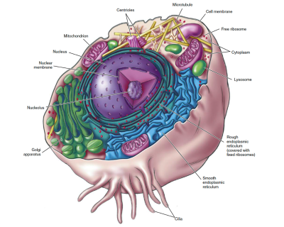
[Alt-Text] A 3-D illustration of a cross section of a cell revealing its interior components. Each part is labelled. See link below for a long description.
[Long Description] Terms from the labels in the diagram appear within quotation marks throughout the description.
The cell has a thin, peach coloured, spherical outer “Cell Membrane”, with a cluster of hair-like roots called “Cilia” projecting from the bottom. The cell is cut in half revealing the inner parts.
In the center is the yolk-like nucleus, which includes:
- “Nuclear Membrane”: A dark purple skin like covering around the Nucleus. It is full of large pores.
- “Nucleolus”: the core of the nucleus; surrounded by a darker, bumpy, sphere.
- Encircling the “Nucleus” is the smooth turquoise ribbon-like “Rough endoplasmic reticulum (covered with fixed ribosomes.)” The Rough endoplasmic reticulum is made up of a series of connected flattened sacs that are part of a continuous membrane that folds to create thin layers around the Nucleus.
The “ribosomes” are depicted as dozens of small red dots stuck to the Rough endoplasmic reticulum. A few are scattered throughout the rest of the cell and are called “Free ribosome”.
Surrounding the Rough endoplasmic reticulum are the other various parts of the cell. Starting at the top and moving clockwise they are as follows:
- “Microtubule”: narrow, yellow, hollow tubes that criss-cross over each other with a web-like substance connecting them. They take up the top third of the cell.
- “Cytoplasm”: the fluid inside the cell, but outside the nucleus.
- “Lysosome”: small pink bean shapes found throughout the cell.
- “Smooth endoplasmic reticulum”: large blue ribbon like structure with many small folds. It takes up a full side of the cell.
- “Golgi apparatus”: a stack of flat sacs that appear like a wide folded green ribbon. It takes up a fourth of the bottom side of the cell.
- “Mitochondrion”: pink slipper shaped structure with two layers. The outer layer is smooth, and the inner layer has many folds. They are located throughout the cell.
This next example is from the same webinar, and is a good example of a top to bottom breakdown.
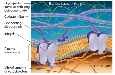
[Alt-Text] A labelled diagram of the extracellular matrix of an animal cell. See link below for a long description.
[Long Description] The labelled sections of the ECM are as follows from top to bottom:
- “Extracellular Fluid”: fills the space outside the cell above the plasma membrane.
- “Glycoprotein complex with long polysaccharide”: long thin green string with dozens of delicate feather like appendages. The strings weave and wrap around the Collagen Fiber
- “Collagen Fiber”: Thicker smooth purple striped strings that float across the extracellular fluid
- “Connecting glycoprotein”: Small purple worm like structures that link the Collagen Fiber to the Integrins
- “Integrins”: double purple bean like shapes clustered together and embedded in the Plasma Membrane
- “Plasma membrane”: the outer membrane of the cell. Described in Figure 4.2.
- “Microfilaments of cytoskeleton”: sets of twisted orange two-strand strings that connect to the plasma membrane
- “Cytoplasm”: fluid that fills the space within the cell below the plasma membrane.
This next example is from the same webinar. It features an insert image, and is a good example of leaving out some details to focus on the main parts of the image.
[Alt-Text] Labelled diagram of plant cell walls with a zoomed in diagram of the plasmodesmata (a channel between the cells). See the link below for a long description.
[Long Description] Plant cells stick together in a honey-comb pattern. The center of each cell has a clear sac like area labelled: “Vacuole”. The plant cell walls are beige and thick with a thinner and darker outer membrane.
The plasmodesmata (channels) are depicted all around the cell walls, as holes in the outer walls of the cells, and as small tunnels between the connected cells.
The zoomed in area shows the details of a Plasmodesmata in the cellular wall. It is labelled from top to bottom as follows:
- “Pectin layer between cells”: is a dark vein in the center of the cellular wall.
- “Primary cell wall”: is located on either side of the Pectin Layer
- “Secondary cell wall”: is located on either side of the Primary Cell wall and is three times thicker.
- “Plasmodesmata”: a tube like channel that passes through the wall from one cell to the other.
- “Plasma membrane”: is located around the Secondary Cell Wall and is a thin darker layer.
- “Cytosol”: liquid inside the cell that can move through the channels.
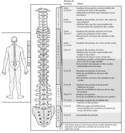
[Alt Text] A diagram titled “Effets d’une lesion médullaire” of the human spinal column. The diagram is divided into three parts. The first part shows the spine, divided into four segments, in relation to the human body. The second part shows the individual vertebrae of the spine, divided into spine segments, labeled with the vertebrae names. The third part is a list of injury locations along the spine and the resulting effects. See the link below the image for an extended description.
[Long Description] A diagram titled “Effets d’une lesion médullaire” divided into three parts.
The first part shows the spine column superimposed over a human body. A legend indicates shading of the four spine segments, labeled with their first letter: C, T, L, and S, and the approximate length of each segment in relation to one another:
- C stands for the Cervical segment and extends the length of the neck.
- T stands for the Thoracique segment and is the longest section that extends from the base of the neck to the mid-lower back.
- L stands for the Lombaire segment and extends the length of the lower back to the top of the hip bones.
- S stands for the Sacre segment and extends the length between the hip bones down to the tail bone.
The second part is a closeup of the spinal column showing the individual vertebrae divided into spine segments. Each vertebra is labeled with its segment letter and ordered numbers as follows:
- The Cervical section includes seven vertebrae: C1 through C7. The C1 vertebrae is disc-shaped and wider than the other vertebrate.
- The Thoracique section includes 12 vertebrae: T1 through T12. These vertebrae are slightly wider and thicker than the vertebrae in the cervical segment, with T1 and T2 being more disc-shaped and wider than the other vertebrae in this section. The vertebrae gradually narrow further down the spine. Each of the Thoracique vertebrae have protrusions on either side representing the ribcage .
- The Lombaire region includes five vertebrae: L1 through L5. These vertebrae are wider and thicker than the Thoracique region.
- The Sacre region encompasses five fused vertebrae: S1 through S5. The region is shaped like an upside-down triangle with blunt corners and small holes on each side of the central vertebrae.
The third part of the diagram breaks down the effects of spinal cord injuries per section as presented in the table below:
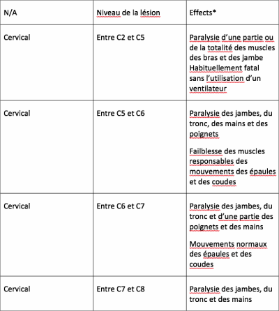
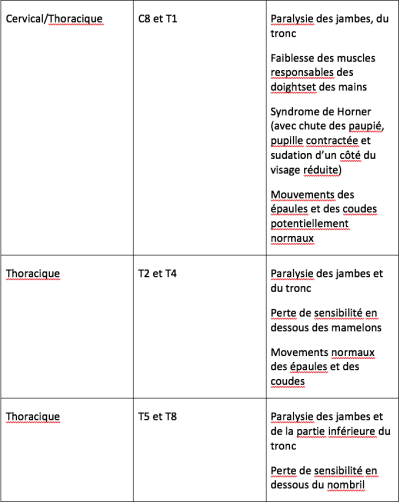
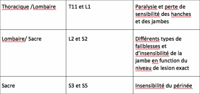
*Note for Effects: Une lesion grave à tout niveau de la moelle épiniére peut entrainer une perte du contrôle de la vessie et du sphincter rectal.
The next 2 examples is from the DAISY Webinar Art and Science of Describing Images Part 3 and is written by Huw Alexander from textBOX
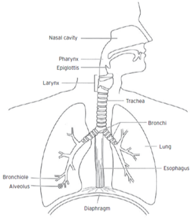
[Alt-text:]A diagram illustrates the main components of the human respiratory system. See the link below the image for an extended description.
[Long Description:] The diagram is a line drawing of the torso and head of a human. The head is turned and faces to the right. The organs of the respiratory system are viewed in cross-section. The respiratory tract is divided into upper and lower sections, as follows:
Upper Respiratory Tract.
- Nasal Cavity. The nasal cavity is a large, air-filled space above and behind the nose in the middle of the face.
- Pharynx. The pharynx is the part of the throat behind the mouth and nasal cavity. It is positioned above the esophagus and larynx.
- Epiglottis. The epiglottis is a leaf-shaped flap in the throat that prevents food from entering the trachea and the lungs.
- Larynx. The larynx, or voice box, is a triangular shaped organ in the upper region of the neck. It is situated just below where the tract of the pharynx splits into the trachea and the esophagus.
Lower Respiratory Tract.
- Trachea. The trachea, or windpipe, is a tube of cartilage that connects the larynx to the bronchi of the lungs. The trachea is around 1.8 centimeters in diameter.
- Bronchi. The tubes of right and left main bronchi enter the lungs. They are around 1 to 1.4 centimeters in diameter.
- Lung. The lungs are found in the chest on either side of the heart in the rib cage. They are conical in shape. The upper lung tapers to a narrow, rounded apex. The broad concave base of the lung sits on the convex surface of the diaphragm.
- Bronchiole. The bronchioles are the smaller branches of the bronchial airways in the lungs. Bronchioles are approximately 1 millimeter in diameter.
- Alveolus. The alveoli are air sacs at the end of the bronchioles in the lungs. These tiny, balloon-shaped air sacs are arranged in clusters, like grapes, throughout the lungs.
- Esophagus. The esophagus is an organ through which food passes from the pharynx to the stomach. The esophagus is a fibromuscular tube, about 25 centimeters long in adults, which travels behind the trachea and heart, passes through the diaphragm and empties into the uppermost region of the stomach.
- Diaphragm. The diaphragm is a sheet of internal skeletal muscle that extends across the bottom of the thoracic cavity. It separates the thoracic cavity, containing the heart and lungs, from the abdominal cavity. As the diaphragm contracts, the volume of the thoracic cavity increases, creating a negative pressure within, which draws air into the lungs.
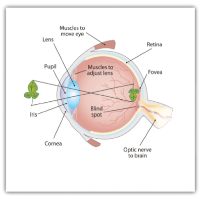
[Alt-text] A diagram illustrates the anatomy of a human eye. See the link below the image for an extended description.
[Long Description]
The eye is viewed in cross-section. The eye can be divided into 2 segments, the anterior and the posterior. The components of the 2 segments are as follows.
- Anterior Segment, representing the front third of the eye.
- Cornea. The outer layer of the eye is controlled by muscles.
- Pupil. The pupil is the aperture or hole in the centre of the iris.
- Iris. The iris surrounds the pupil.
- Lens. The lens is an ellipsoid structure that sits behind the pupil and iris and refracts light into the retina. Muscles control the adjustment of the lens.
- Posterior Segment, representing the rear two thirds of the eye.
- Cavity. A cavity containing a clear gel.
- Retina. The retina is the innermost layer of the eye and connects directly to the optic nerve.
- Fovea. The fovea is a small depression or pit within the retinal surface.
- Blind spot. The blind spot is an area on the retinal surface that does not contain photo receptors.
The optic nerve runs from the back of the eye to the brain.
The diagram illustrates how the retina interprets an image as upside down and backwards. In the example, a leaf is used to illustrate this concept.
